
Preoperative Localization
When a lumpectomy is planned, we need to be able to remove the cancer with a small amount of surrounding tissue. If the cancer is not palpable — was detected by mammography, ultrasound or MRI — the surgeon needs a guide to know where to go to find the cancer. The radiologist provides this guide or "roadmap" for the surgeon, by placing either a magnetic seed or a wire in the breast using mammography and/or ultrasound.
About the Procedures
Preoperative Magnetic Seed Localization
VCU Health is the first in Richmond to offer preoperative localizations with a magnetic seed - called Magseed. This is scheduled as an outpatient procedure prior to the day of surgery at either the Nelson Clinic or Stony Point.
Similar to your biopsy, the magnetic seed is placed in the tumor under local anesthesia using mammographic or ultrasound guidance. You can report directly to Pre-Op on the day of Surgery, and go home the same day. While you are in the operating room, the surgeon uses a special camera or detector to locate the magnetic seed and tumor in your breast.
The tissue that the surgeon removes will be brought back to Breast Imaging and we will x-ray it to confirm removal of the tumor and the Magnetic seed. We mark the location of both in the tissue specimen so the pathologist knows where to focus their attention in analyzing the specimen.
Preoperative Localization
On the day of your surgery you will be asked to go to Room 300 of the Nelson Clinic (on the third floor, across from the elevators) so that a small wire can be placed through the cancer using mammographic or ultrasound guidance. The wire stays in your breast long enough to serve as a road map for the surgeon to find and remove the cancer.
After the wire is placed in your breast, you are transported to the surgical waiting area in a wheelchair.
While you are still in the operating room, the tissue that is removed will be brought back to Breast Imaging and an X-ray image is done to confirm that the abnormality has been removed from your breast. We will also make sure that the wire used to localize the lesion has been removed from your breast and we mark the location of the abnormality in the specimen so the pathologist knows where they need to focus their attention in analyzing the specimen.
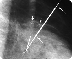 In this patient, there is a cluster of microcalcifications (small single arrows) in the left breast. A wire (long single arrows) with a small hook (double arrow) at the end is placed through the cluster of calcifications. The hook serves to keep the wire in position. The tissue that is removed is X-rayed to verify that the lesion — in this patient, calcifications — is excised. The X-ray is also used to make sure that the wire comes out of the breast and to tell the pathologist where to focus their attention.
Everyone I came in contact with was absolutely wonderful to me — the VCU Medical Center is lucky to have you all on staff.
- Excerpt from a VCU Breast Imaging patient satisfaction survey.