Innovative VCU research lab reopens with scanner upgrade and new avenues for brain and metabolic imaging
The Collaborative Advanced Research Imaging facility — VCU's research-dedicated MRI site at the Wright Center — returns from a comprehensive renovation.
August 28, 2026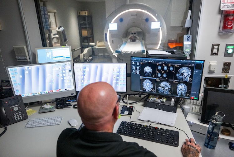 Robert Cadrain, MRI manager and chief technologist at the Collaborative Advanced Research Imaging facility, views images from CARI’s new Philips MR7700 3T MRI system, which brings dramatically expanded research possibilities for investigators. (Dean Hoffmeyer, Enterprise Marketing and Communications)
By Christopher Richmond
One of Virginia Commonwealth University’s most unique facilities — a research site that has supported studies spanning addiction, neurology, maternal health, genetics and adolescent development — has reopened after a months-long renovation that installed a star attraction: a new scanner that opens new windows into human biology.
CARI, the Collaborative Advanced Research Imaging facility, has been a cornerstone of VCU’s brain imaging enterprise for more than a decade. Administered by the C. Kenneth and Dianne Wright Center for Clinical and Translational Research, CARI is currently home to more than 60 active studies, and its research-dedicated MRI — calibrated specifically for scientific protocols rather than clinical throughput — gives investigators access to technology that most universities share with busy hospital imaging departments.
The renovation of CARI’s 6,000-square-foot building on East Cary Street includes a new Philips MR7700 3T MRI system, which brings dramatically expanded research possibilities for investigators across VCU and beyond.
But “the entire building went through a transformation,” said Robert Cadrain, MRI manager and chief technologist at CARI. “No part of the CARI facility was untouched,” from its basement HVAC room to the rooftop chiller that keeps the scanner magnet cold.
The MR7700 replaces CARI’s previous Philips Ingenia scanner, and the technical gains are substantial: They include a 44% increase in gradient strength and improved heat management, which allows the system to sustain demanding scan protocols without slowdown or overheating. For fMRI specifically, the new system can acquire 20% more brain volumes in the same scan time.
Renovated facility and new scanner will advance VCU research
Wright Center Director F. Gerard Moeller, M.D., professor and addictions division chief in VCU’s School of Medicine and director of VCU’s Institute for Drug and Alcohol Studies, has built a large research program around CARI. That includes VCU serving as one of 21 sites participating in the Adolescent Brain Cognitive Development Study, the nation’s largest-ever longitudinal neuroimaging study of human brain development.
CARI is where the brain scans for VCU’s ABCD participants, including more than 550 children and 215 pairs of twins, take place.
“The new system builds on that legacy while opening doors that were not previously available to VCU researchers,” Moeller said.
The scanner upgrade brings meaningful benefits to VCU’s participation in the ABCD Study. The MR7700’s enhanced gradient performance allows VCU’s diffusion MRI protocols to align with parameters used at other ABCD sites nationwide.
“This infrastructure update ensures our neuroimaging data meets contemporary structural and functional research standards, particularly for our ongoing projects in addiction and adolescent brain development,” said Joel L. Steinberg, M.D., CARI’s medical director and a research professor in the School of Medicine.
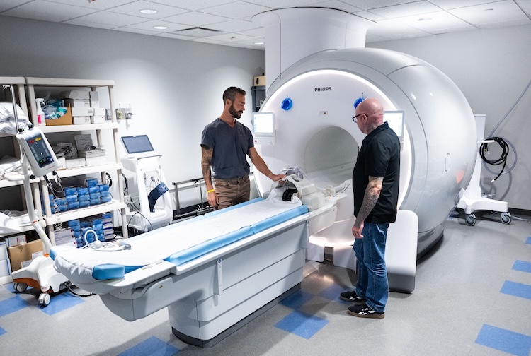 Eric McFadden and Robert Cadrain with the Philips MR7700 3T MRI system. (Dean Hoffmeyer, Enterprise Marketing and Communications)
Brain and metabolic imaging at VCU: New frontiers await exploration
Perhaps the most consequential change is what the new scanner can detect that the old one could not.
The MR7700 introduces multinuclei imaging and spectroscopy – the ability to image not just hydrogen (the basis of conventional MRI) but also phosphorus, carbon, sodium, fluorine and xenon – without requiring specialized external coils. Each opens a different window into the body’s biology.
- Phosphorus spectroscopy allows researchers to watch muscle metabolism unfold in real time – for example, tracking mitochondrial function during exercise recovery.
- Carbon-13 imaging can map metabolic processes in the liver and heart – this can include how diabetes medications alter glucose production.
- Sodium imaging can detect early signs of stroke, tumor response and cartilage degradation.
- Xenon gas inhalation enables functional lung imaging without radiation – this can help with mapping ventilation and assessing conditions such as COPD, asthma and cystic fibrosis.
The system also introduces two noncontrast imaging techniques valuable for patients who cannot tolerate traditional contrast agents. 3D Amide Protein Transfer imaging may help distinguish tumor progression from treatment effects, and 4D-TRANCE provides detailed views of blood vessels in patients with kidney disease.
CARI serves both the Monroe Park and MCV campuses and is open to investigators from across VCU as well as external partners. In addition to scanner access, the facility offers a mock MRI, physical exam space with an ECG machine, and expert consultation for protocol design and data analysis. With the addition of a second MRI technologist, the facility offers extended weekday hours through 8 p.m. and newly available weekend slots. Investigators interested in using CARI can email cctr_cari@vcu.edu or visit go.vcu.edu/cari.