Dr. Thomas Porter Presents Research
Dr. Thomas Porter has given new meaning to the term “scrubbing bubbles.”
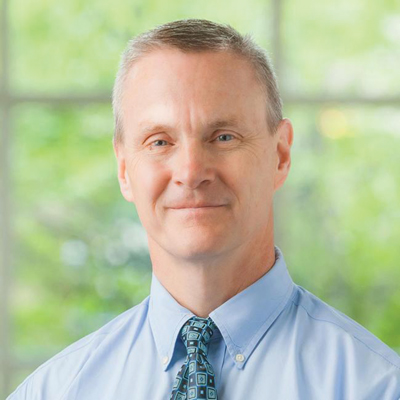
Porter, a former resident and cardiology fellow at the Medical College of Virginia, who is now the Theodore F. Hubbard Endowed Chair of Cardiology at the University of Nebraska Medical Center (UNMC), has spent many years studying the efficacy of using microscopic bubbles known as “microbubbles” to break up blood clots. Although the microbubbles are used primarily for imaging, they can also be used to break up a blood clot using the same process used for ultrasound imaging. He presented his most recent findings from a clinical trial at the American College of Cardiology meeting in March.
“It was the culmination of the first 100 patients we had tested, [exploring] whether this process would work in patients having an acute heart attack,” he said by phone, from his office in Omaha, this past April.
The study focused on patients in Brazil who were experiencing a heart attack. Before undergoing cardiac catheterization, they were first injected with commercially available microbubbles through an IV. During the short time period before reaching the catheterization laboratory, they underwent a diagnostic and therapeutic ultrasound.
“We demonstrated that the procedure improved the blood flow in the major blood vessels causing the heart attack, but more importantly, improved the blood flow to the microvasculature,” said Porter. “It also helped the heart recover from the heart attack, whereas it wouldn’t have recovered if we’d just done a stent.”
His interest in microbubbles began in 1990, when he was a cardiology fellow at MCV. At that time, he was exploring the use of the microbubbles as an ultrasound enhancing agent for imaging, work that he continued when he joined the faculty of UNMC in 1992.
“Microbubbles are smaller than your red blood cells, they travel with your red blood cells, and they are bright reflectors of sound. We get beautiful images with ultrasound using them,” he said.
Over a period of 20 years, from early test tube explorations to those involving animal models, he discovered that the process of imaging causes the bubbles to shake–a process known as cavitation.
He explained that, as a bubble grows and collapses, “it creates a bit of a micro-tsunami around it, and that little tsunami, when it’s next to a blood clot, breaks it up without the need for Heparin or other clot-breaking agents.” As a result, “we can avoid bleeding complications, which is very advantageous. We think that this treatment will work not only for acute heart attacks but for acute strokes as well.”
Additionally, the procedure may also help prevent the onset of heart failure that typically occurs in patients with an acute myocardial infarction who fail to recover the full pumping function of the heart after stent placement. The sound waves from the imaging not only break up blood clots in the large vessels but those in the microvasculature—”all the tiny blood clots that just shower down to your smaller blood vessels during a heart attack that cause you to develop scars and cause your heart to be weakened following a heart attack.”
“One of the secondary findings in the Brazil trial was that the number of patients that needed to have a defibrillator at six months following a heart attack was cut by 75%,” he added.
He plans to soon initiate a study in Amsterdam, where ambulance technicians will inject the microbubbles and apply ultrasound to patients who they have identified as having a heart attack. Recent advancements in technology allow the technicians to perform the ultrasound imaging on laptop-sized machines.
“It’s a very safe process, and the earlier you get it started, the more likely you expect to see the blood clot break up, not only in the patient’s large blood vessel but in the microvasculature,” said Porter.
The next step will be a multi-national ambulance trial, and hopefully, FDA approval in the U.S. will follow.
“We’re very excited about this,” he said. “For probably the better part of 20-25 years, we’ve been trying to find ways to address this problem of the scarring and damage that occurs downstream in a heart attack. A lot of research has gone into this. But this is really one of the first treatments that’s been tested in humans that actually works.”
Memories of MCV
After completing his medical degree at University of Nebraska Medical Center, Dr. Thomas Porter spent many years of his medical training at MCV, including a residency in internal medicine from 1984-1988 (he served as chief resident from 1987-1998) and a cardiology fellowship from 1988-1991. He served on faculty the year following.
His interest in microbubbles first began during his fellowship, when one of his mentors, Dr. George Vetrovec, asked a compelling question: “`Find a way to help me figure out whether a blockage is causing a patient’s symptoms. If you can find a way to do that, it would be so helpful,’” Porter recalled, with a laugh.
“At that time, there was no real way we could, at the bedside, assess whether a blockage in the coronary artery was affecting the blood flow to the patient’s heart muscle, and whether it was causing their chest pain,” he explained. “We subsequently were able to invent microbubbles and ultrasound imaging techniques that would allow us to do that both at rest and during stress testing. Today, this ultrasound imaging technique is done around the world. So hopefully we have answered Dr. Vetrovec’s question.”
In addition to Vetrovec, Dr. Kenneth Ellenbogen, Dr. Ian Nixon and Dr. Walter Paulson were important teachers. Of the latter, Porter said, “he took so much time to teach you the technique of echocardiography. And no one studied the literature better than Walter Paulson. Whenever I was on any kind of rotation during my cardiology fellowship, I would sneak out of the rotation just go over to the echo lab and listen to him discuss cases as he was reviewing echos for the day because you would learn so much.”
He recalled his first impressions of the Richmond hospital as a resident. “I went from a small medical center in Omaha to this huge, developing, exciting place where there were patients everywhere and all kinds of illnesses that I’d never seen before,” he said. As he began the long rotations—all while raising a young family–“every month was just a tremendous learning experience. The hardest days of your life, but the best days.”
Porter returned to Richmond in 2011, when he was named VCU Alumni of the Year. He spoke at both the medical school and cardiology grand rounds. While much had changed in terms of the hospital’s growth and new technologies, “the interest and the enthusiasm of the faculty had remained the same. All those great faculty have really been the fabric that has made Virginia Commonwealth University such a special place to be.”
Back to Autumn-2019