New Cardiovascular Imaging Suite Debuts
“The new Cardiovascular Imaging Suite was designed with multidisciplinary collaboration in mind,” said Dr. Greg Hundley.
Filled with numerous monitors and screens displaying MRI and cardiopulmonary exercise test (CPET) findings, the suite’s 435-square-foot control room provides space for Hundley to work with a wide array of experts in cardiovascular medicine and radiology as well as biomedical engineering, medical informatics, physical medicine and rehabilitation, and behavioral medicine. The group is exploring heart failure in cancer and cardiac patients.
“The new suite is a cutting-edge diagnostic unit offering a combination of equipment, procedures and expertise only available at a few top-tier institutions,” said exercise physiologist Justin Canada, Ph.D.
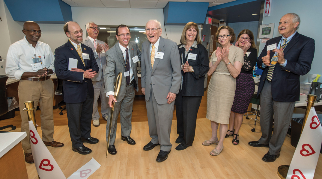
A $4 million donation by Stan and Dorothy Pauley and family made the suite and Hundley’s recruitment and research possible, and enabled Pauley to receive an additional $5 million gift for the project from the Glasgow Endowment. Stan Pauley and his daughter, Kathy Pauley Hickok, attended the official ribbon-cutting held July 17.
“Our visit to the new suite was most gratifying. We are very excited by Dr. Hundley’s vision and are pleased to have been able to support this unique endeavor,” said Stan Pauley. “We think the work that will be conducted here will have a major impact on the lives of many heart and cancer patients.”
Located on the ground floor of the North Hospital, the 7,200-square-foot suite is decorated in warm earth tones. Blue-accented entrances guide patients from waiting to changing rooms, then on to rooms for procedures and recovery. A plexiglass wall with rows of 2-inch holes allows for improved communication between the patient recovery and control rooms.
A sign indicates “Magnet Always On” at the doorway to the room that holds a new Siemens Magnetom Vida 3T MRI system, which just received FDA approval last fall.
“MRI CPET imaging is done by only a few centers in the world,” said exercise physiologist Justin Canada, Ph.D.
The term “3T” refers to a 3 Tesla magnet, indicating the strength of the magnetic field. The new Siemens wide bore MRI is the first 3T system of its kind at VCU Health and generates a field twice as strong as the existing 1.5T machines. The 3T runs faster, is less vulnerable to background noise, and produces higher resolution images as a result. The new Biomatrix Sensors of the Siemens’ model help anticipate a patient’s breath-holding capability, to reduce the percentage of poor quality scans.
“It’s the Cadillac of the 3 Tesla systems,” said Brent Metts, MRI modality manager at VCU’s Massey Cancer Center.
Heart studies involve more than 1,000 images and can take 45 minutes to an hour to complete at many facilities. “With the new equipment, for some patients, we can complete the studies in as little as 15 minutes,” said Hundley, who explored the effectiveness of these shorter MRI sessions as principal investigator of a multi-center, NIH-funded study of patients undergoing the imaging for the treatment of cancer.
The new suite offers electrocardiogram (ECG) stress testing, stress echocardiography, exercise MRI, and MRI CPET testing for patients. Normally, patients who undergo CPET have a catheter placed in their wrist, to draw blood samples during the test; breathe into a tube to measure ventilation, oxygen consumption and carbon dioxide production; and are hooked up to a blood pressure cuff, pulse oximeter and ECG leads. The new testing will include all these measurements coupled with magnetic resonance imaging. A bicycle attachment will allow patients to pedal to get their heart rates up while lying inside the MRI.
“MRI CPET imaging is done by only a few centers in the world,” said Canada. “It will refine our ability to detect early signs of cardiopulmonary disease and more accurately determine the causes of exercise intolerance in symptomatic patients.”
According to Dr. Ann Fulcher, chair of the Department of Radiology, “the lab will enhance collaboration between radiologists and cardiologists as exercise/stress testing followed by magnetic resonance imaging will be performed in the same area.” She added, “this will promote a close working relationship as these physicians strive to provide optimal diagnostic examinations for their patients which can then be used to guide therapy.”
The suite provides technologies and opportunities not available at any other academic institution in Virginia, North Carolina or Maryland and will draw other VCU Health experts and students as well as from VCU’s School of Engineering and other academic programs, said Hundley.
“This is going to be a training facility for the next generation,” he said.
FROM L TO R: DR. VIGNESHWAR KASIRAJAN, DR. KENNETH ELLENBOGEN, DR. PETER BUCKLEY, DR. GREG HUNDLEY, STAN PAULEY, KATHY PAULEY HICKOK, DR. MARSHA RAPPLEY, DEBORAH DAVIS, AND DR. GORDON GINDER CELEBRATE THE RIBBON-CUTTING OF THE NEW CARDIOVASCULAR IMAGING SUITE.
Back to Autumn-2018
Join our Pauley Consortium composed of patients, friends and advocates.