Coming Home
Pauley Welcomes Dr. Greg Hundley as First Director
It’s April, and Dr. Greg Hundley, VCU Health Pauley Heart Center’s first-ever director, has started to move into his new office in West Hospital. Around the room are stacks of boxes filled with books. A dry-erase board, not yet hung, leans against a wall.
He wears a dark suit and a stylish tie. He is 56 years old but looks years younger.
His glasses give him the appearance of a scholar—which, indeed, he is. Among his achievements, he has more than 200 peer-reviewed articles and $24 million in NIH grants, and 19 years of continuous NIH funding to his name. Despite this, Hundley is approachable. Sitting across from a visitor at a round table near his desk, he is friendly and eager to talk about his new role with Pauley.
“There’s an enormous opportunity here with the talent at VCU Health. We have expertise in heart failure, cancer treatment, electrophysiology, image acquisition and analysis, bioinformatics, biomedical engineering and coronary heart disease,” he said. “We have renowned physicians like Dr. Kenneth Ellenbogen; a phenomenal CEO of the hospital, Deborah Davis; and an exciting new dean with a great vision, Dr. Peter Buckley.”
“I was very impressed with Dr. Hundley when I first met with him due to several factors, among which are his very humble and pleasant personality, his excellent reputation as a physician-scientist, as well as his vision for the heart center,” said Dr. Fadi Salloum.
While winding down his practice at Wake Forest Baptist Medical Center, where he served as program director for the cardiovascular imaging program, he helped the MCV Foundation secure funds to build a new $4 million Cardiovascular Imaging Suite at VCU Medical Center with the magnetic resonance imaging (MRI) technology necessary to do his high-caliber work. The foundation raised an additional $5 million for Hundley’s recruitment and research.
“It was an amazingly generous gift of Stan and Dorothy Pauley and their family, in their tradition of commitment to excellence in health care delivery, that made this all possible,” said Hundley.
The Pauleys’ gift enabled the foundation to receive an additional $5 million in matching funds from the Glasgow Endowment.
The suite will support his groundbreaking work in preventing and treating heart failure in cancer patients and mark a new era of collaboration between Pauley and VCU’s Massey Cancer Center.
“Massey has been one of the research sites with which we have collaborated in the past for my trials,” he said. “Now, with the new lab, I’m going to work side by side with these same physicians as well as VCU engineering and VCU Health exercise physiologists, behavioral scientists and cardiovascular medicine specialists. We are going to be using the very latest MRI and cardiopulmonary exercise test technologies, which are going to open up all kinds of possibilities. I’m looking forward to seeing what these collaborations will bring.”
“We are all very excited to have him join and lead Pauley,” said Dr. Antonio Abbate, associate chair of research. “Hundley is an internationally renowned expert in cardiac imaging. He is not only a skilled clinician but also a talented researcher. He also brings an extensive experience in mentoring trainees and junior faculty.”

“I was very impressed with Dr. Hundley when I first met with him due to several factors, among which are his very humble and pleasant personality, his excellent reputation as a physician-scientist, as well as his vision for the heart center,” said Dr. Fadi Salloum, Natalie N. and John R. Congdon Sr. Endowed Chair.
When asked about the impact of hiring Pauley’s first director, Ellenbogen said, “It is highly significant and very exciting. It will be like taking us from the major leagues to the World Series.” He added, “Dr. Hundley is going to provide us with the depth in research and cutting-edge clinical research that will attract physicians from all over the world to study, train and do research with us.”
Hundley grew up in Richmond, then attended the College of William and Mary, where he met his wife, Kim, a native of Ashland. After graduating in 1984, he attended medical school at VCU School of Medicine.
“I was here when the transition occurred to move the main clinical operation from West Hospital to Main Hospital,” he said. During his long hours as a medical student, he remembers taking a tunnel to the capitol to eat at Chicken’s. Skull and Bones was another favorite.
“My class was large, with a lot of diversity and talent and phenomenal clinical and my desired research training,” he said. “Research-wise, I had the distinct opportunity to work with Dr. Hermes Kontos as well as Drs. Joseph Levasseur, Enoch Wei and Joe Patterson.”
Hundley felt drawn to cardiovascular medicine after working in the cardiovascular ICU. “The patients were relatively sick, and there was a lot of satisfaction in the care that could be delivered in that environment to make them well.”
From 1988 to 1996, he completed his internship and residency in internal medicine, as well as his fellowship in cardiovascular disease at Parkland Memorial Hospital and the University of Texas Southwestern (UTSW) Medical Center in Dallas. There, while originally training to be an interventional cardiologist, he grew fascinated by cardiac imaging.
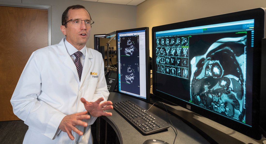
His world changed the first time he saw an MRI. Parkland Hospital was one of few facilities in the country to own the advanced imaging system in 1993. “One morning I went over to pick up a patient from the magnetic resonance imaging scanners who was scheduled to undergo a heart catheterization. On the screen of the MRI scanning console was an image of a beating heart as if it was being displayed on a high-definition TV,” he said. “Thirty years ago, that was an amazing accomplishment.”
Today, as during the 1990s, the cardiac catheterization lab used invasive procedures incorporating angiograms—x-ray photos obtained by using contrast dye injected through catheters placed in the arteries of one’s arms or legs—to detect blockages in the coronary arteries. At Parkland and UTSW, investigators were working on noninvasive MRI methods that did not require an interventional heart catheterization procedure to visualize and measure blood flow in these same coronary arteries. However, the process of viewing the heart and these arteries took a long time. “It took probably an hour and a half to take 10 pictures of the heart, and then you stayed up all night typing binary commands so you could see them the next day,” he recalled. “Literally all night, until 4 to 6 a.m.”
Today, instead of all-night sessions, “with MRI you can acquire and visualize a complete 3D image data set of the whole heart in 15 seconds,” he said with a laugh, still amazed.
His world changed the first time he saw an MRI. Parkland Hospital was one of few facilities in the country to own the advanced imaging system in 1993.
Inspired, he became involved in research with mentors Drs. David Hillis, Richard Lange, Geoff Clarke and Ron Peshock to corroborate MRI findings by comparing them to coronary angiograms. The research, published by the American Heart Association, gained significant international attention. He also devised protocols for the use of MRIs in creating images of coronary arteries that supplied the heart muscle.
During this time, his innovative work caught the attention of Dr. George Vetrovec, the former chair of cardiology who retired in 2015. “We sat next to each other at a dinner of the Society for Cardiac Angiography and Intervention. He had just won a prize from the organization for imaging studies,” recalled Vetrovec, who later tried to recruit Hundley—twice—but the timing wasn’t right.
Vetrovec recalled his first impressions. “I thought he was very, very intelligent and well-spoken and thoughtful. He had this innovative new technology and was at the forefront of its development.”
While Hundley liked Dallas, he and his wife longed to get back to the East Coast, closer to family. When Dr. William Little at Wake Forest Baptist Medical Center offered him a place on the faculty in 1996, he accepted.
“They recruited me there because they had a lot of patients experiencing heart attacks and they wanted to use this new MRI technology clinically,” he said.
Hundley and Little further developed clinical MRI protocols to image coronary arteries and measure blood flow within them in patients presenting with chest pain suspected of experiencing a heart attack. At the time, patients with blocked arteries were treated with intravenous thrombolytic therapy, which involved using clot-busting drugs. With this therapy, “we could never tell whether the infarct artery was open or closed, and this MRI technique could do this.”
After he’d been there less than three months, a superior new technology emerged that replaced thrombolytic therapy: balloon angioplasty. This meant MRI was not needed to view the coronary arteries because patients went straight to the heart catheterization lab to not only view the arteries but directly open them.
“So, I was stuck. Because I’d moved there to introduce this new technique, and Little came in said, `Well, we’re not going to do that anymore.’ I said, `What am I going to do?’ And he said, `Well, you’ll think of something.’”
He soon discovered that being a physician deep in the heart of tobacco and barbecue country presented special challenges. “A lot of people had coronary artery disease, but they were not very good candidates, because of their smoking and obesity, to have pictures made of their heart noninvasively to determine if they needed a heart catheterization.” Noninvasive echocardiography and radioisotope techniques were often inadequate in these patients.
Hundley, along with Drs. Craig Hamilton and Kerry Link, began using an MRI to image their hearts, “and that really took off, because it provided an answer for a population that didn’t have one.” A story about their noninvasive MRI ran on NBC Nightly News.
Next, he began to grow interested in heart failure while doing stress tests. While these tests are usually done on just the heart, he and Little began testing all the blood vessels in the body. “Heart failure is a complex disease where not just the heart has problems contracting or relaxing, but also the blood vessels that carry the blood have problems,” he said. “With MRI, one could image the heart, blood vessels, kidneys and other structures simultaneously. In so doing, one could determine why tissues weren’t receiving the blood supply they needed.”
Hundley, in collaboration with Drs. David Herrington and Dalane Kitzman at Wake Forest, studied the etiologies of heart failure when the heart ejection fraction is preserved, or shows up as normal, on echocardiograms. This latter syndrome, common in older individuals, often causes heart failure to go unidentified until it is significantly progressed.
His interests took a turn one day, while working with program manager Kim Lane and acquiring a MRI of a patient who’d had heart failure after being treated for cancer. “The type of imaging we were doing always produced a picture that was black or white,” he said. “But this time we got gray.”
The physicians first blamed it on a glitch in the new software program. However, they later realized that the gray areas indicated early heart damage from the cancer treatments. Often this damage could be detected before permanent injury occurred.
“The chemotherapies and the radiation therapies were actually damaging the heart and blood vessels in addition to treating the cancer,” he said. “The heart has a high metabolism, so it makes sense that it would be susceptible.”
Working with his associates, Hundley received more than $20 million in NIH funding for a research trial to use MRIs to identify patients who were developing heart injury from their cancer treatment. Additionally, he has had the opportunity to work with a number of emerging younger clinicians and scientists in studying mechanisms and methods to prevent heart attacks, strokes, heart failure and exercise intolerance in those treated for cancer—later putting them into clinical practice.
“Oncologists are so effective in treating cancer that now, for many cancers, heart problems are the primary cause of the death for these patients,” he said.
With all these discoveries, Hundley was highly recruited by other facilities. The opportunity to work at his alma mater as the first-ever director, with a state-of-the-art imaging suite, proved irresistible.
Hundley will be able to continue and expand his work in the new Cardiovascular Imaging Suite. The new 7,200-square-foot imaging suite includes capabilities to assess tissue anatomy, perfusion, function and metabolism with high temporal resolution at rest and during exercise. VCU Medical Center will be one of the few sites in the world capable of these tests.
Although his work focuses on cancer patients, “the techniques are applicable for all patients with cardiovascular disease,” he said. Because of cancer’s rapid metabolism rate, the disease provides a special window to understanding heart disease.
“High blood pressure causes heart or vessel damage, but it takes 60 years for you to see that, so that’s not great for conducting a study. But in a cancer patient, because of how quickly the disease metabolizes, that 60 years is shrunk into weeks,” he said.
“The developments we’re finding in the cancer patient population will help us better understand heart and vascular disease and high blood pressure, diabetes, patients with high cholesterol and all the other causes of heart failure,” he said.
“It’s enormously exciting for us to be able to do this in central Virginia.”
To get a preview of the new Cardiovascular Imaging Suite, please see “New Cardiovascular Imaging Suite Debuts” on page 14.
About Dr. Hundley
“Dr. Hundley is a nationally and internationally recognized leader in his area of research on the cardiac complications of cancer therapy. His ability to collaborate with faculty at VCU Massey Cancer Center is already demonstrated through our participation in his cardiac toxicity prevention trials, which bodes well for productive future collaborations between our two centers. We are excited about him joining us at VCU Health.”
— Dr. Gordon Ginder
Director, VCU Massey Cancer Center
“Dr. Hundley is a proud alumnus of VCU School of Medicine, and we are equally proud of him. With his outstanding reputation as a physician-scientist and mentor, Hundley is a perfect fit as medical director. His research in the exciting program of cardio-oncology will build on our strengths. This is central to our strategy to attain comprehensive cancer center designation for Massey. Additionally, Dr. Hundley will elevate Pauley’s national prominence and federal research portfolio.” — Dr. Peter Buckley
Dean, VCU School of Medicine
Did you know…
- Since 1999, Dr. Hundley has been involved with more than $71 million in awarded research funded grants.
- He currently serves as associate editor for Circulation and Cardiology Today and serves on the editorial board of five other peer-reviewed journals.
- He is married to his loving wife of 30 years, Kim Hundley, a speech pathologist, “who has encouraged and tirelessly supported my career,” he said. The Hundleys have three children: Anna, 22; Jennifer, 18; and Will, 17.
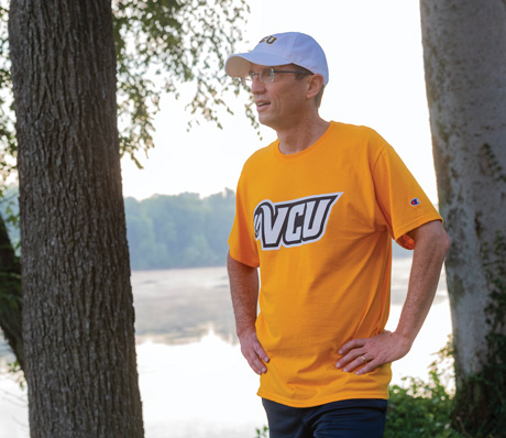
FROM TOP TO BOTTOM: DR. GREG HUNDLEY POINTS OUT THE SUPERIOR IMAGING OF THE SIEMENS MAGNETOM VIDA 3T MRI SYSTEM. DR. GREG HUNDLEY RUNS ALONG THE JAMES RIVER. HE IS A LONGTIME ADVOCATE FOR THE IMPORTANCE OF ADOPTING A HEALTHY LIFESTYLE.
Back to Autumn-2018
Join our Pauley Consortium composed of patients, friends and advocates.